Cryo-EM: A New Tool to Help Researchers Develop Therapeutics Against Cancer
Cancer is a disease where a group of cells no longer behave normally, and instead grow out of control and spread (or metastasize) to other parts of the body. It sounds simple, but the truth is that it is far more complex [and explained more in depth in a previous post]. Scientists have been trying to disentangle how cancer functions and survives for decades. It has been a difficult road, but important advances have been made. However, to keep progressing, the complexity of cancer demands that different scientific approaches and disciplines band together to drive new discoveries and new treatments.
In recent years, an up-and-coming approach has risen out of the field of structural biology and proven to be quite useful in cancer research (and many other fields). This method is called cryo-electron microscopy (cryo-EM) and is such a powerful tool that the group of scientists that developed it received the Nobel prize in chemistry in 2017. So what is cryo-EM and how is it used? Briefly put, it is a method where a beam of electrons is shot at a frozen protein sample to capture an image of the protein’s 3D structure. Scientists can then analyze the protein structure to reveal key information about its function and how it interacts with other proteins.
Knowing the structure and function of proteins is a key aspect to understanding cancer biology (and why the field of structural oncology has formed). This is because proteins with altered structure and function due to genetic mutations are what drive cancer development. So if scientists can understand how a protein’s structure is changing from genetic mutations by using cryo-EM, they may learn about how a protein is triggering the development of cancer. Cryo-EM can also reveal how other proteins and molecules (like drugs) can bind to a specific protein, which can assist in identifying a way to target a cancerous protein with a therapeutic agent.
Other powerful imaging methods have also existed and been used to identify protein structures, such as x-ray crystallography and NMR spectroscopy. This leads to the question, what makes cryo-EM special? Although x-ray crystallography and NMR spectroscopy are excellent methods in their own right, they also pose certain caveats that cryo-EM does not.
For example, In x-ray crystallography, the proteins have to be crystallized for viewing, however, not all proteins are able to be crystallized and some proteins change their structure when they are. Cryo-EM does not require crystallization. For NMR spectroscopy, it is usually restricted to smaller proteins or parts of a protein, whereas cryo-EM is not. Cryo-EM also has the capability of viewing proteins that are embedded in cell membranes, form complexes with other proteins, and can view proteins moving and interacting as they perform functions. So overall, cryo-EM has immense potential to be a powerful tool.
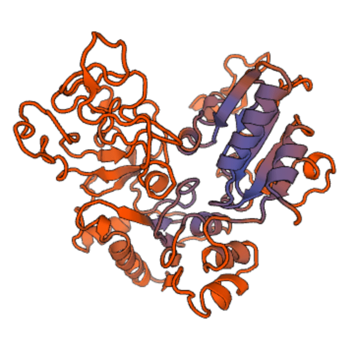
An example of cryo-EM at work can be seen in a recent study published in Science Advances. This study focused on the protein encoded by the oncogene (or gene that can cause cancer when mutated) called USP1 because its gene expression is upregulated (or turned up) in several types of cancers – including breast cancer, ovarian cancer, colorectal cancers, and bone cancers – often with poor prognosis. This makes USP1 a promising drug target in some contexts. Additionally, inhibitors of USP1 have been shown to decrease the growth of cancer cells. Surprisingly though, how inhibitors bind to this protein has been unknown. That is why this recent study aimed to characterize the binding of inhibitors to this protein using cryo-EM.
Using cryo-EM, the scientists achieved a high resolution view of the molecular binding of the well-established inhibitor of USP1 (called ML323). They did this by studying the molecular structure of USP1 with and without the inhibitor bound to it, which revealed an unusual binding mechanism. Rather than binding to a surface pocket, the inhibitor binds to the hydrophobic (or ‘water hating’) core of the protein. This binding by the inhibitor disrupts the catalytic site (or the part of the protein that performs an action) of USP1 to inhibit function. Having this new understanding of how an inhibitor actually binds to USP1 is an important advancement that can aid scientists in developing new inhibitors and treatments against cancers where USP1 is upregulated.
.jpg)
That is a general theme occurring with the use of cryo-EM – scientists are learning the exact mechanism of how proteins function and how drugs bind to them to help guide improvements on existing treatments and the development of new ones. This includes other studies using cryo-EM, such as a study on the cancer drug rituximab, a study on the cancer target IDH, and multiple studies on the mutations of the oncogene PI3Kα and its conformational changes during activation and inhibition. So it is safe to say cryo-EM use will only continue to rise. This is reflected by the fact that the number of protein structures determined by it continues to grow rapidly, as shown by the rising submissions to the Electron Microscopy Data Bank [EMDB – a repository for protein structure].
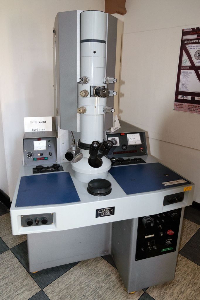
However, there is concern that the cost of the method could be a hindering factor for some scientists. Obtaining a capable microscope can cost >$7 million (USD); preparing a room suitable for the scope and installing it can cost a similar amount; and it can cost an upwards of $10,000 (USD) a day to run it. Some scientists can have the option to network and send out samples to a location that has a scope already, or pay for the service, but wait times can be painfully long ranging from 3 months and beyond. Then, by the time one has access, more time is added because cryo-EM can be a difficult process to get correct with many trial and errors. Additionally, many structural biologists do not have access at all. That is why some scientists in the field are trying to design a machine that can do the same method at a much lower cost. Hopefully advances in the method/machine can make it more affordable and accessible to more scientists.
Overall, this new technology has the potential to make it easier to create more individualized and protein-specific treatments that will uniquely target cancer causing proteins. We may even be able to discover ways to target proteins that were believed to be completely ‘untargetable‘. This should inspire hope as we continue to make the steps in the right direction, and perhaps one day cryo-EM will help scientists develop the ‘silver bullets’ against cancer we all dream for.


Leave a comment